Yeatman, a research scientist at I-LABS, studies how the brain learns to read. While a graduate student in Brian Wandell’s lab in the Stanford University Department of Psychology, he was examining MRI images of human brains when he noticed a prominent fiber bundle that seemed to be a part of the intricate network of brain connections that process visual information.
But when Yeatman tried to look up the pathway in reference books, he couldn't find it. And so began some scientific detective work to solve the mystery of the "lost highway," as he calls it.
In a paper published Nov. 17 by the Proceedings of the National Academy of Sciences, Yeatman and his colleagues at Stanford describe how scientific confusion and controversy dating back to the late 1800s led to the fiber path being left out of the neuroscience literature. They also provide software to help other researchers identify the brain structure in their own data.
Read more about the findings and see historical images of the brain in a university news release.
Yeatman did the work with Wandell and Kevin Weiner, a postdoctoral researcher at Stanford University.. It was a side project for all of them, so as time permitted, they would head down to the basement of Stanford Medical Library and explore old brain atlases – some of which only a few copies exist in the world. Over a few years and a half-dozen or so visits to the library, they pieced together the story of the lost highway – known now as the vertical occipital fasciculus.
Here they talk more about what they found and what it may mean:
What prompted you to do this study?
Yeatman: I wanted to know how a major piece of brain anatomy could get forgotten and essentially written out of the scientific literature. I first noticed the vertical occipital fasciculus a few years ago when I was examining fiber-tracking results from diffusion MRI scans of human brains. As the name implies, diffusion MRI measures the rate of water diffusion in the brain and allows us to trace bundles of nerve fibers that carry information between distant brain regions. I was amazed by a massive fiber bundle going into a region of cortex called the visual word-form area. This region of the occipital lobe is critical for skilled reading. I couldn't find this pathway in any brain atlas, so my adviser Brian Wandell sent our figure to some colleagues in neurosurgery to see if they knew of it. Eventually, one of them replied saying he recalled seeing something like this in an old medical textbook. So we started tracing its history through other neuroscience references from the turn of the 20th century.
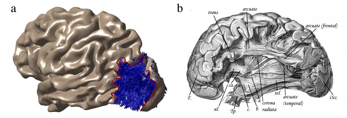
The figure below is a side by side comparison of a side view of the brain with the occipital lobe – where visual information is processed – pointing toward the right. The image on the left depicts the vertical occipital fasciculus as a bundle of blue fibers in the occipital lobe. It is taken from Yeatman and colleagues' diffusion MRI measurements. On the right is a drawing of the same region of the brain by neuroanatomist Theodor Meynert. It appears in am article published in 1892 and while it points out other prominent brain pathways, it leaves out the vertical occipital fasciculus – which would be at the far right side of this illustration, behind the layers of cortex. By covering up the VOF with cortex, "It's like Meynert was saying 'Nothing to see here,'" Yeatman joked. The VOF contradicted Meynert’s theory that all association fibers traveled from back to front rather than vertically.

What was it like going through old scientific texts?
Weiner: Perhaps it's a shock to hear that not everything is online. But, not everything is online yet. I think very few students these days know what it's like to check your belongings at the door when you sign in to a rare books section of the library. And there is something magical about going through that process and sitting in front of atlas after atlas - of smelling the ink and turning page after page. There are so many amazing details within historical texts that can really motivate present hypotheses and even potentially resolve scientific debates. For Jason, Brian, and I, as well as our co-authors, this was a real motivating factor – to search through these old texts to find what hints these historical giants of neuroscience had provided for us as to why this important white matter bundle in our brain has been forgotten for so long.
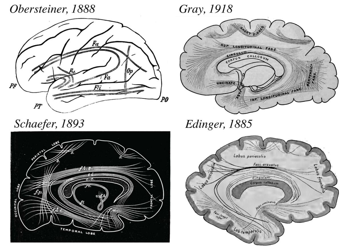
The series of images below shows drawings of brain connections that the researchers found in the various atlases they studied. Heinrich Obersteiner's 1888 camera lucida drawing (considered state-of-the-art method at the time), Sir Edward Schaefer's 1893 woodblock carving, and Gray's 1918 illustration all show the vertical occipital fasciculus. But Ludwig Edinger's 1885 drawing leaves out the fiber pathway.

What was the highlight for you?
Yeatman: The "aha" moment for me was when Kevin flipped to a page in an atlas written by Carl Wernicke in the late 1890s. An image depicted a pathway in the occipital lobe that was oriented vertically, crossing the other major fiber tracts that connect the back and the front of the brain. We knew right away that this was the vertical occipital fasciculus or "senkrechte Occiptalbündel" as Wernicke named it.
Weiner: Probably the most memorable moment was turning a page in an atlas, by Schnopfhagen and finding out that the evidence leading the charge against the existence of the VOF was from the brain of a calf embryo. We all had a good laugh about that and it really reminds you how much the field has advanced from the days when just having any brain would suffice to make what was at the time, a logical argument.
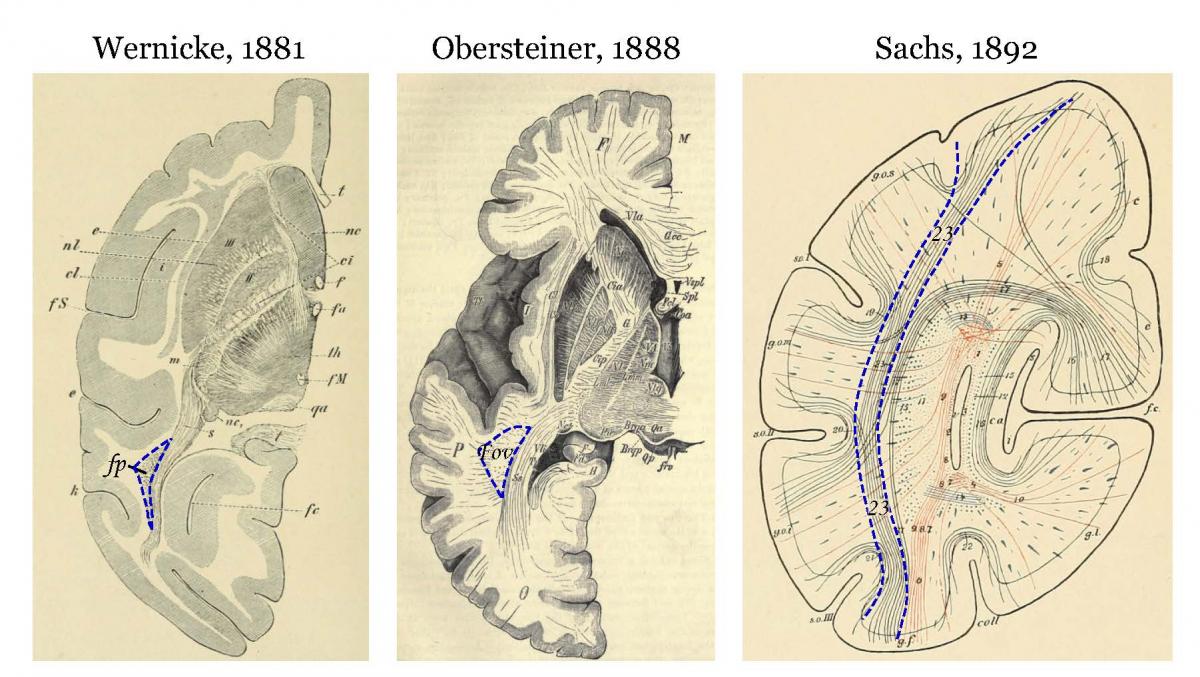
Below are the first images of the vertical occipital fasciculus, with varying names and abbreviations. Seeing Wernicke's 1881 drawing from a monkey brain was the "aha" moment that helped the researchers piece the story together. The drawings by Obersteiner and Sachs are from human brains.

What does a calf embryo have to do with human brains?
Yeatman: That’s what made it so funny! We kept reading in different texts that Franz Schnopfhagen was arguing with Wernicke and his student Heinrich Sachs about the existence of the VOF. Apparently these attacks bothered Sachs enough that he devoted a few paragraphs of a publication to defending his and Wernicke’s discoveries against Schnopfhagen's attacks. We would have never guessed that the disagreement centered on comparing human anatomy to that of a calf embryo. I’m not quite sure how it would even be possible to identify the same structures in each brain.
What's next?
Weiner: By publishing these findings, we hope to not only preserve a bit of history, but also to replicate images of postmortem histological sections from over 100 years ago and display them alongside brain images taken with non-invasive, contemporary methods in live brains. We're hopeful that future studies will uncover all the different contributions that the VOF makes to different aspects of human vision and cognition.
Yeatman: I'm excited that we now have a detailed picture of the anatomy of the VOF and can move toward understanding how the signals carried by this pathway contribute to human cognition. The question that keeps me fascinated by neuroscience is how the brain learns to read. And the VOF may be a significant player in that process. There were a couple of papers in the 1970s showing that injuries to the VOF produced profound reading impairments. Now that we have much more precise ways to measure the brain non-invasively, we have the opportunity to follow up on these findings in a number of ways. I’m beginning to work on a study that follows children with dyslexia through an intensive reading intervention program to see how their brains change as their reading skills improve. I wouldn’t be surprised if the VOF, once again, ends up at the center of this story.
Selected Media Coverage
The Guardian
Washington Post
KUOW
Smithsonian
i09
LiveScience
Seattle PI
The Scientist
Fox News
UPI
###